Home

Laboratory

Wet Lab Techniques

  • Cell Culture
  • Gel Electrophoresis
  • Pipetting and Solution Preparation
  • Aseptic Technique

Dry Lab Techniques

  • PyMOL
  • Swiss-Model
  • CB-Dock2
  • Chimera
  • BLAST
  • NCBI/UniProt/PDB

Analytical Methods

  • Spectrophotometry
  • Chromatography
  • Hemocytometer

Imaging

  • Fluorescence Microscopy
  • Fiji (ImageJ)
  • Xenon MRI
  • CT Segmentation

Cell Staining & Imaging
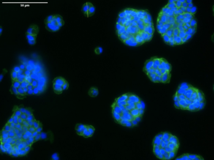
Nuclei Fluorescence Microscopy
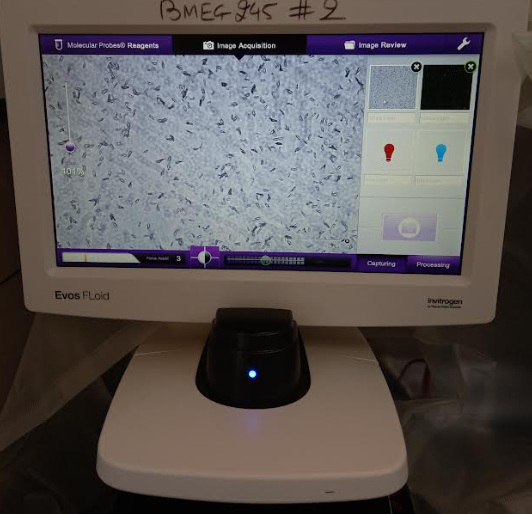
White Light Microscopy
Green Light Microscopy

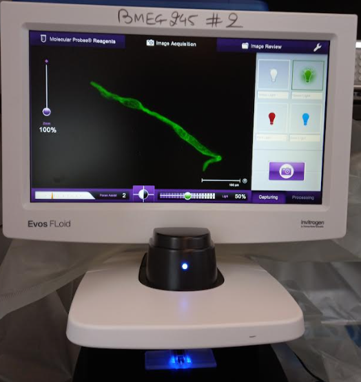
Problem: Visualizing intracellular structures is critical to understanding cell morphology and organization. I wanted to observe nuclei and actin filaments in mammalian cells to learn fluorescence imaging techniques and study cell structure.

Solution: I prepared cell cultures in an IBIDI imaging chamber, fixed them with paraformaldehyde, permeabilized with Triton X-100, and blocked nonspecific binding using BSA. Cells were stained with phalloidin Alexa-Fluor-488 and Hoechst dye to label F-actin and nuclei. Using fluorescence microscopy, I captured images based on photon emission and wavelength patterns.

Impact: This project taught me hands-on skills in fluorescence staining, microscopy, and image analysis. I gained experience in aseptic techniques, cell culture preparation, and using fluorescent markers to study intracellular structures.

Features: Fluorescence Microscopy, Image Processing, Cell Imaging, Fiji (ImageJ), Aseptic Technique, Micro Pipetting, Cell Culture
DNA Manipulation

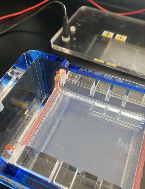
Gel Electrophoresis Setup
DNA Ladder
DNA Ladder

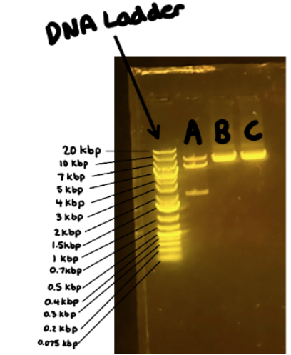
Problem: Separating DNA fragments is essential for cloning and genetic analysis. I wanted to learn how plasmid DNA can be manipulated and visualized to prepare for downstream applications like cloning or amplification.

Solution: I digested plasmid DNA with restriction enzymes in an optimized buffer, producing DNA fragments of different sizes. Using agarose gel electrophoresis, I separated the fragments based on size, with a conductive buffer enabling the electric field. This allowed visualization and isolation of specific fragments for potential cloning into host cells like E. coli.

Impact: This project taught me hands-on skills in plasmid manipulation, enzymatic digestion, gel setup, and DNA visualization. I gained experience with precise micro-pipetting, understanding DNA charge properties, and the workflow for preparing DNA for cloning experiments.

Features: Gel Electrophoresis, DNA Manipulation, Restriction Enzymes, Agarose Gel, DNA Ladder, Micro Pipetting

Protein Engineering

3EQM Structure
Human Aromatase Structure
3EQM Active Site
Human Aromatase Active Site

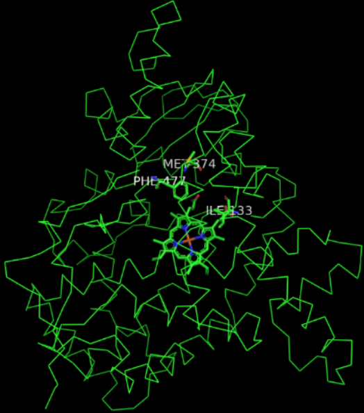
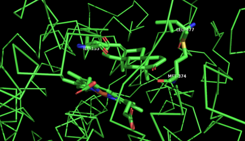
Problem: Low estrogen levels can lead to osteoporosis by weakening bone structure. I wanted to study the aromatase enzyme, which converts testosterone and androstenedione to estradiol and estrone, and explore how mutations could optimize its activity to improve estrogen production.

Solution: I analyzed the enzyme's active site, identifying three key residues in the binding cavity. I designed mutations to adjust steric and chemical properties: choosing bulkier, aromatic residues to reduce pocket size and maintain hydrophobic compatibility with the target molecules. I then updated the FASTA sequence in Swiss-Model to generate mutant enzyme models and used CB-Dock 2 to dock ligands and estimate Gibbs free energy for binding feasibility.

Impact: This project strengthened my skills in molecular modeling, enzyme active site analysis, and computational docking. I gained experience evaluating structural and chemical compatibility for protein engineering and predicting the effects of mutations on enzyme activity.

Features: Molecular Modeling, Active Site Analysis, PyMOL, Swiss-Model, CB-Dock 2

Protein Release Kinetics

Protein Assay
Protein Assay
Protein Assay Demo
Protein Assay Demo

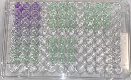
Problem: Understanding how proteins are released from hydrogels is essential for designing controlled drug delivery systems. I wanted to study the kinetics of protein diffusion and release over time to evaluate material performance.

Solution: I immersed hydrogel spheres in buffer and collected samples at specific time intervals. Protein concentration was measured using a BCA assay, with absorbance quantified via spectrophotometry and compared against a standard curve to generate release profiles and calculate diffusion rates.

Impact: This project helped me develop hands-on skills in protein quantification, spectrophotometry, and reaction kinetics. I learned to analyze release profiles to assess diffusion behavior and gain insight into material performance for biomedical applications.

Features: BCA Protein Assay, Spectrophotometry, Chromatography, Reaction Kinetics